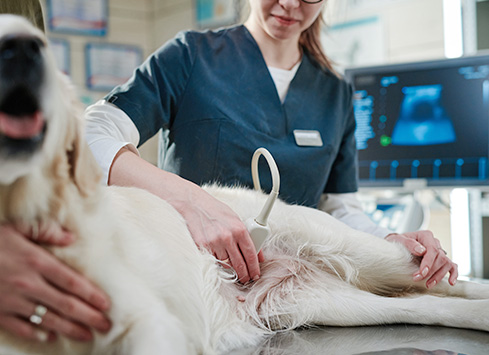
Ultrasound machines produce still and dynamic images using soundwaves.
Ultrasound is non-invasive and particularly useful for visualising blood flow and organs. Images are stored on our PACS system and can be viewed on any of our IRIS workstations throughout the practice.
Common uses for ultrasound:
General
On occasion, when a patient is not stable enough for a CT or X-ray, ultrasound can be used to check for pathologies, including pleural effusions or bladder stones. It is often a good test for looking at the liver architecture and other organs.
Heart scans
Ultrasound enables viewing of the heart in multiple imaging planes. We can visualise the four chambers of the heart and the major vessels. It is particularly useful when investigating cardiac disease, including heart murmurs, arrhythmias and ECG changes.
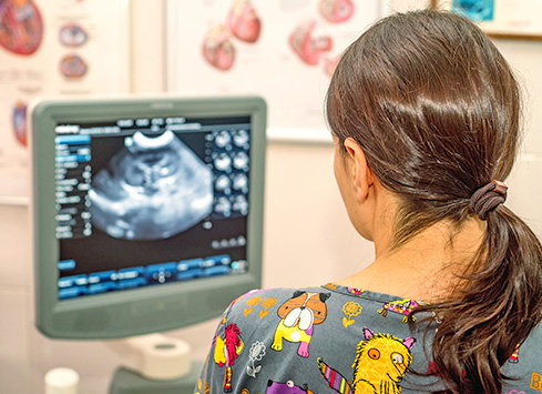
Pregnancy scanning
Ultrasound is not harmful to an unborn foetus and is the modality of choice to confirm pregnancy. Uterine disorders, including pyometra, may be diagnosed using ultrasound.
Biopsy and drainage guidance
Ultrasound guides the operator during these procedures and leads to more accurate results than blind sampling. It allows for a more precise placement of the biopsy needle or drain, avoiding other structures.
Doppler colour flow
This mode of scanning is used to determine the presence and direction of blood flow. It can help to establish whether there are any profound circulation abnormalities.
Tumour detection
If a mass is suspected, ultrasound can be used to distinguish any focal lesions from surrounding structures.
Cystocentesis
If a non-contaminated urine sample is needed, a sterile needle can be guided into the bladder using ultrasound control, and a sample can be obtained.