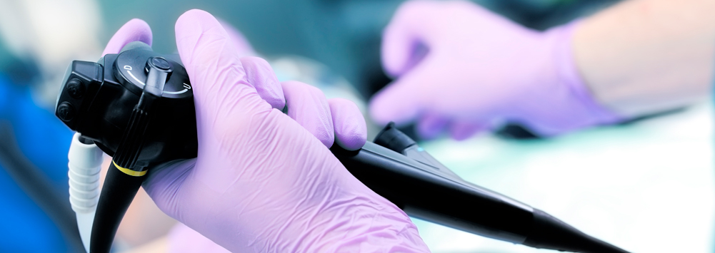
Endoscopy allows for the examination of an animal’s internal organs, via a live stream. The technique uses a flexible or rigid device, dependent upon the area of the body being observed. Images from the camera are transmitted, in colour, onto a screen, guiding the operator and allowing them to identify pathologies.
Common sites for endoscopic examination are the trachea, lungs, oesophagus, stomach, bowel, nose, bladder, ears, joints and rectum. Endoscopic procedures are relatively quick and non-invasive. More often than not, the patient is anaesthetised to avoid any discomfort. Endoscopy is a valuable tool for examining inside the body and is key when taking biopsy samples from internal organs.
Common uses for endoscopy:
Endoscopy of the respiratory tract can involve one of two different procedures, dependent upon the symptoms displayed by the animal.
Rhinoscopy: this is a procedure used to examine the nose and back of the throat. Rhinoscopy can be used to investigate sneezing, nasal discharge or bleeding from the nose. It is particularly useful when looking for foreign bodies and to investigate nasal congestion.
Bronchoscopy: during this procedure, a scope is passed down into the upper airways and lungs. Bronchoscopy can be used to investigate a chronic cough, respiratory issues, pneumonia or airway disease. In addition, biopsies can be taken and a bronchoalveolar lavage (BAL) can be performed. A BAL is the introduction of a sterile saline solution into the airways, which is then suctioned back out. The fluid can be looked at cytologically to assess the types of cells present and can also be cultured, to detect any bacterial or fungal growth.
Common pathologies seen:
- Tracheal collapse
- Foreign bodies
- Masses
- Inflammation
- Aspergilloma
- Nasal polyps
- Fungal infections
A variety of gastric pathologies can be diagnosed using different gastroscopes and a range of procedures.
A gastroscope is the scope used when looking at the oesophagus, stomach and bowel. In order for the bowel lumen to be viewed, food is normally withheld from patients, for a short time. Using the scope, biopsies can be taken and strictures can be dilated.
Oesophagoscopy: is a procedure used to visualise the oesophagus, checking for any strictures, foreign bodies or growths. Clinical indications include difficulty swallowing, excessive salivation or drooling and regurgitation.
Gastroscopy: is a procedure used to look for any bleeding, polyps or masses in the stomach. Typical indications include vomiting, weight loss, anorexia and loss of appetite.
Duodenoscopy: allows vets to examine the duodenum. The major duodenal papilla resides inside the duodenum and provides the entrance to the common bile and pancreatic duct. A scope enables vets to view strictures and masses, and to assess the papilla, in patients suffering from chronic diarrhoea, vomiting, anorexia and weight loss.
Colonoscopy: requires patients to have a series of enemas or to take a ‘Klean Prep’ solution, emptying their bowels completely. Clean, empty bowels are essential for clear visualisation. A flexible scope is passed, via the anus, to look at the colon. Clinical indications for this test include the presence of fresh blood in faeces, diarrhoea or difficulty in passing solids. This test can help us to rule out any tumours or intestinal bleeding.
Common pathologies seen:
- Gastric ulcers
- Foreign bodies
- Oesophagitis
- Strictures
- Polyps
- Neoplasia
- Diverticulum
- Hernia
- Intussusception
Otoscopy is the name used for the endoscopic procedure that examines the inside of an ear.
A rigid scope is used to pass through the external ear into the external ear canal. Foreign bodies, including grass seeds, commonly make their way into animal’s ears and can cause irritation or infection. Clinical indications for an otoscopy include pain, discharge, scratching or rubbing the ears, shaking of the head and sometimes a head tilt.
Common pathologies seen include:
- Tumours
- Foreign bodies
- Otitis externa
- Otitis media
Arthroscopy is the procedure through which the interior of a joint is examined.
Arthroscopy: this technique uses a rigid scope to gain direct visualisation of the synovial cavity, inside a joint. This method causes minimal disruption to the joint structures and less scar tissue, meaning patients recover much faster than if they were to undergo a surgical procedure.
It can be used as a diagnostic and therapeutic tool, to investigate and treat joint conditions, including:
- Elbow disease
- Shoulder instability
- Osteochondritis disseccans
- Osteochondrosis
- Meniscal injuries
- Cruciate ligament tears
- Fragmented coronoid process
- Ununited anconeal process
- Septic arthritis
- Biciptal tendon disease
Arthroscopies can be used to remove small bone chips and fragments from inside a joint, as well as to assess damage to articular cartilage and to take biopsies of the synovial membrane. It is possible to work on a number of joints during one procedure, reducing the need for multiple anaesthetics. Pictures and videos can be captured, in order to document findings and show clients.

