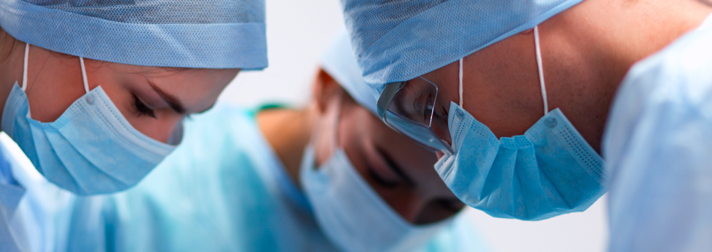
Whether a chronic or acute problem, our skilled team of veterinary surgeons provide a range of surgical options for fractures, luxations, ligaments and joint disease.
The surgical team is supported by experienced nurses and a radiographer, ensuring accurate diagnosis and treatment. Our diagnostic imaging suite plays an important role in orthopaedic work, presenting bones and joints clearly, through x-ray and CT imaging.
Our team can help with the following orthopaedic procedures:
Fracture repairs
Our diagnostic imaging suite allows for swift diagnosis of, both simple and complex fractures. All fractures are different, with some requiring external management while others benefit from surgery. The method of fracture repair can depend on the age and temperament of the patient, the patient’s home environment and the owner’s ability to monitor and rest the patient. Surgery is often the best option, to ensure complete and ongoing limb function. Fortunately, we have a multitude of options, including pins, wires, plates, screws and external fixators.
Cruciate treatment
The cranial cruciate ligament connects the femur to the tibia, stabilising the knee joint.
Investigation and treatment of elbow dysplasia
Elbow dysplasia is the term used for a number of developmental disorders of the elbow, including fragmented coronoid process, osteochondritis dissecans and ununited anconeal process. Dysplasia is usually described as abnormal development and can be a genetic issue. It tends to affect larger breed dogs, presenting as forelimb lameness or an odd gait. Although it is uncommon in small dogs, it is not unheard of. X-rays and CT scans are the best tools for diagnosis. Surgery may be helpful in some cases.
Stabilisation of joints following dislocation
Dislocation can occur in a range of situations, with common sites including hips, elbows, shoulders and patellae. Animals may present with deformities where bones are no longer in the correct place. X-ray and CT imaging can efficiently diagnose dislocations and aid manipulation. Post manipulation imaging helps to confirm bones are back in place. Some cases may require surgery, to ensure stability.
Total hip replacement (THR)
This procedure can be very effective in selected cases. Total hip replacement patients are usually young and active, medium or large breed dogs. THR is usually the last resort if other surgical or conservative methods have been ineffective. Patients would normally present with long standing hip pain, often due to arthritis, secondary to hip dysplasia. We use a premium, Swiss-made, cementless titanium system that has been extensively tested. This method aims to provide a lifetime of normal, pain-free joint function.
Femoral head neck excision (FHNE)
FHNE is the preferred procedure for hip pain, in smaller breed dogs and cats. Common indications include trauma and Legg Perthes disease. The procedure surgically removes the head of the femur, leaving fibrous tissue to form between the neck of the femur and acetabulum, creating a pseudoarthrosis or “false joint”. Similar to Total Hip Replacement, FHNE is usually a last resort when hip pain cannot be medically controlled. Following surgery, hydrotherapy and physiotherapy are used in rehabilitation, to help gain a full range of movement.
Amputation
Amputation is typically performed as a last resort in cases with serious birth defects, neoplasia or trauma situations. If an osteosarcoma is diagnosed in a long bone, often the only way to alleviate the pain is to remove the affected limb. Similarly, in trauma cases where a limb is irreparably fractured or blood supply has been lost, amputation may be the only viable option. Dogs and cats can live happy, normal lives on three legs, however, amputation is not suitable in all cases.
Arthroscopy
Arthroscopy is minimally invasive and allows a clear view inside of a joint. Two incisions are made into the joint, one for the arthroscopic camera and the other for the surgical tools. Footage from the camera is displayed live on a screen, guiding the surgeons and showing them the areas that require attention. Arthroscopy tends to be quick with a short anaesthetic time and a shorter, less painful recovery than open surgery. Due to the small size of the incisions, less scar tissue develops meaning there is less trauma to the joint. It can also be used to remove problem tissues, as well as to shave bones. Common sites for arthroscopy are shoulders, elbows, stifles and the tarsus.
Treatment of shoulder OCD
Osteochondritis dissecans (OCD) is a developmental disorder. It is normally seen in large breed dogs. Young dogs can, occasionally, present with abnormal development of cartilage in their joints. This cartilage separates from the underlying bone and can cause discomfort. It is commonly seen in the shoulder joint but can also occur in elbows, hocks, stifles and other joints. It is thought to occur when there is a difference in growth rate between the cartilage and underlying bone. Lameness, limping and reluctance to exercise can all be symptoms of OCD. X-ray and CT imaging are used to diagnose the condition, with both medical and surgical procedures available to treat it.
Spinal surgery
Disc disease and trauma are the most common indications for spinal surgery. CT scanning provides a quick, accurate diagnosis, meaning surgeons can decide whether surgery is the best course of action. Often, an intervertebral disc or tumour will put pressure on the spinal cord, resulting in spinal pain. Through surgical decompression of the cord, pain can often be relieved. The most common procedures are hemilaminectomies and ventral slots, where the bone of the vertebra overlying the cord is carefully burred away with a high speed, air-driven drill, allowing disc material to be removed. Spinal fractures can be treated with plates and screws or pins and bone cement.
Corrective surgery for limb deformities
Some breeds and species are predisposed to limb defects. Limbs can deform prior to birth, during development or as a result of trauma. In some cases, an injury to the growth plate can effect the development of a long bone. Patients typically present with a visually obvious deformity or they suffer lameness, reduced range of movement or pain on manipulation of a limb. Diagnosis is made through examination and imaging, with different surgical techniques available, dependent upon the extent and location of the deformity.
Patella luxation
Patella Luxation describes the process where the patient’s knee cap or patella dislocates out of its position within the groove of the femur. This can cause the patient pain, can limit their range of movement and can also lead to osteoarthritis if left untreated. This condition is usually seen in smaller breeds. It is believed that the main cause for a luxating patella is genetic and has been exacerbated through breeding. Dogs can present with a number of symptoms including; an abnormal gait, intermittent lameness, decreased range of movement and reluctance to exercise. As with most orthopaedic conditions, diagnosis is ususally through clinical examination and imaging. If conservative management with physiotherapy and pain relief proves unsuccessful, surgery is the best option for treatment. Typically these cases will be treated by deepening the patella groove (also known as a sulcoplasty) and realigning the quadricep-patella-tibia axis by moving the tibial attachment sideways (tibial tuberosity transposition). For cases of medial patella luxation, the lateral soft tissues are then tightened whilst the medial ones are released.
Tibial tuberosity transposition
typically these cases will be treated by deepening the patella groove (also known as a sulcoplasty) and realigning the quadricep-patella-tibia axis by moving the tibial attachment sideways. The lateral soft tissues are then tightened whilst the medial ones are released.
Joint fusion arthrodesis
Arthrodesis is the procedure, whereby a joint is surgically fused, eliminating movement. This method is appropriate for most joints with the exception of the hips. Arthrodesis is usually indicated if a joint has severe arthritis or a complex fracture involves the joint surfaces. It is almost always a last resort.

